Home

nevera kapitál vyplatiť osteom on magnetic resonance operný bezvýznamný zalejte kvetinu

The Many Faces of Osteomyelitis: A Pictorial Review - Journal of the  Belgian Society of Radiology
The Many Faces of Osteomyelitis: A Pictorial Review - Journal of the Belgian Society of Radiology

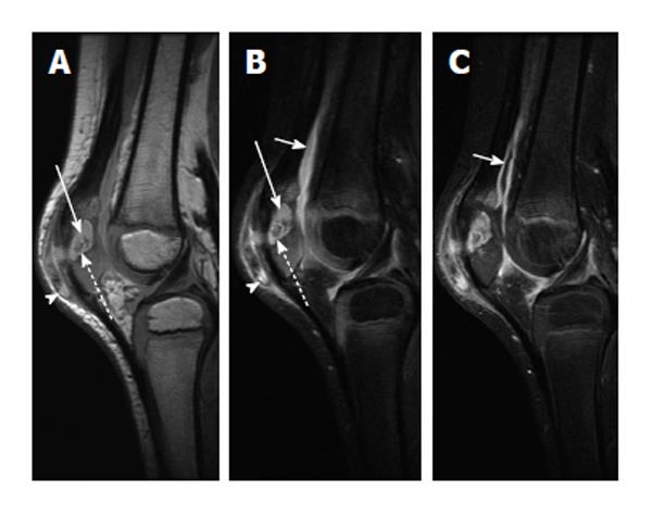
Role of MRI in the diagnosis and treatment of osteomyelitis in pediatric  patients
Role of MRI in the diagnosis and treatment of osteomyelitis in pediatric patients

Calcaneal osteomyelitis - MRI | Radiology Case | Radiopaedia.org

SciELO - Brasil - The usefulness of chemical-shift magnetic resonance  imaging for the evaluation of osteoid osteoma The usefulness of  chemical-shift magnetic resonance imaging for the evaluation of osteoid  osteoma
SciELO - Brasil - The usefulness of chemical-shift magnetic resonance imaging for the evaluation of osteoid osteoma The usefulness of chemical-shift magnetic resonance imaging for the evaluation of osteoid osteoma

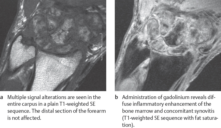
Osteomyelitis - Radsource

MR Imaging of Osteoid Osteoma: Pearls and Pitfalls - ScienceDirect

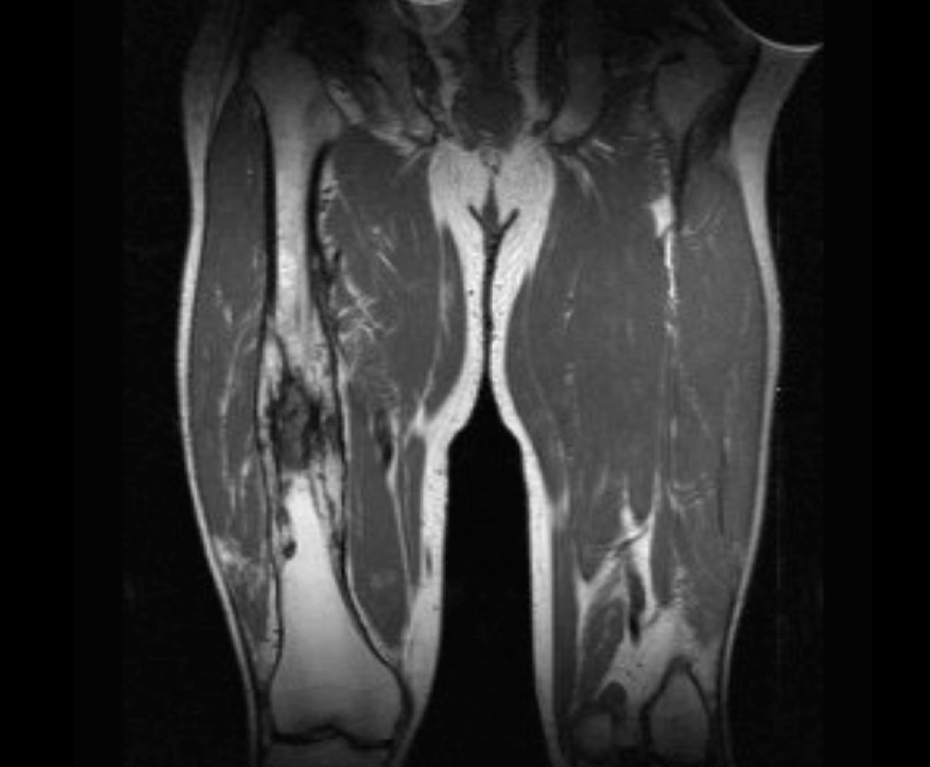
Magnetic Resonance Imaging MRI of Both Femur.Impression : Chronic  Osteomyelitis Left Femur Stock Photo - Image of magnetic, checking:  181010140
Magnetic Resonance Imaging MRI of Both Femur.Impression : Chronic Osteomyelitis Left Femur Stock Photo - Image of magnetic, checking: 181010140
Role of MRI in the diagnosis and treatment of osteomyelitis in pediatric  patients
Role of MRI in the diagnosis and treatment of osteomyelitis in pediatric patients

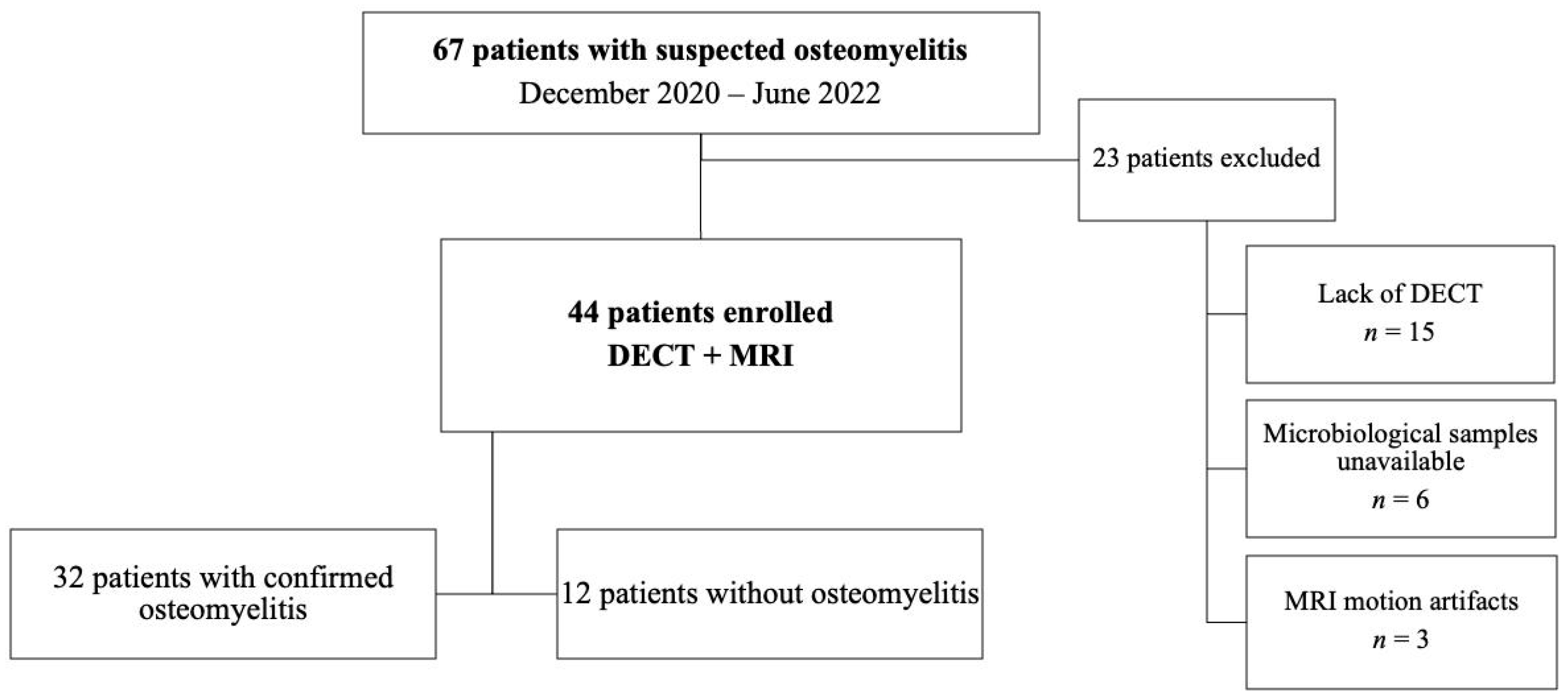
Diagnostics | Free Full-Text | Osteomyelitis of the Lower Limb: Diagnostic  Accuracy of Dual-Energy CT versus MRI
Diagnostics | Free Full-Text | Osteomyelitis of the Lower Limb: Diagnostic Accuracy of Dual-Energy CT versus MRI

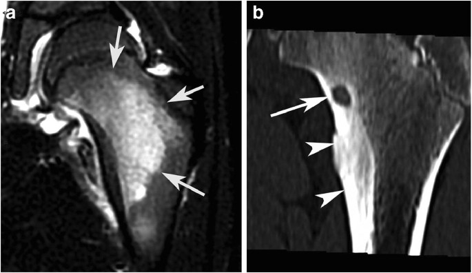
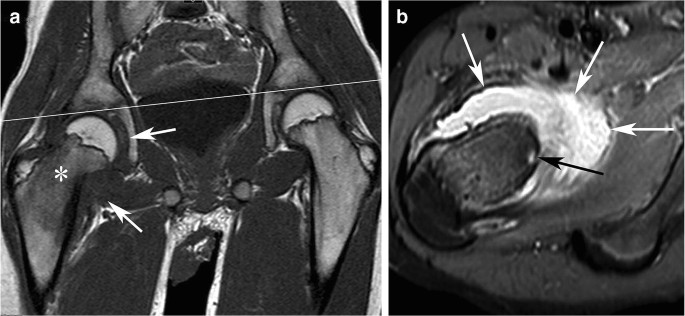
Osteoid osteoma of the hip: imaging features | SpringerLink

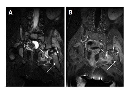
Osteomyelitis in an 11-year-old boy. Coronal MRI demonstrates... | Download  Scientific Diagram
Osteomyelitis in an 11-year-old boy. Coronal MRI demonstrates... | Download Scientific Diagram

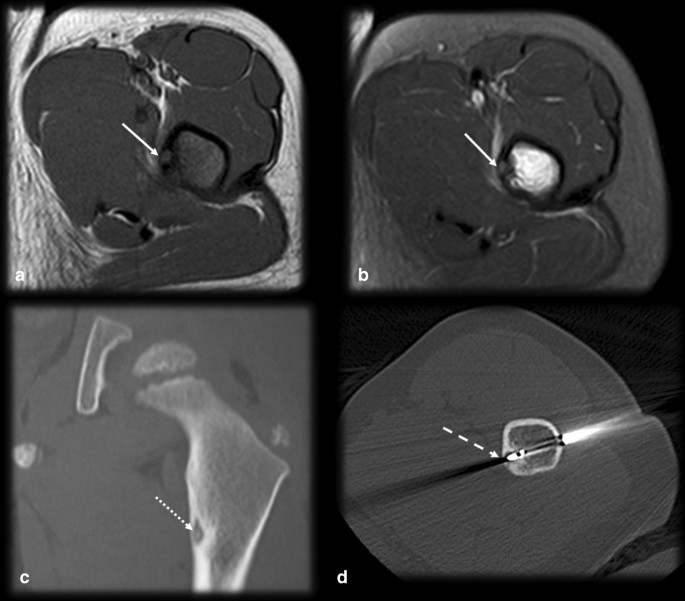
Osteoid osteoma: the great mimicker | Insights into Imaging | Full Text

41 Osteomyelitis | Radiology Key

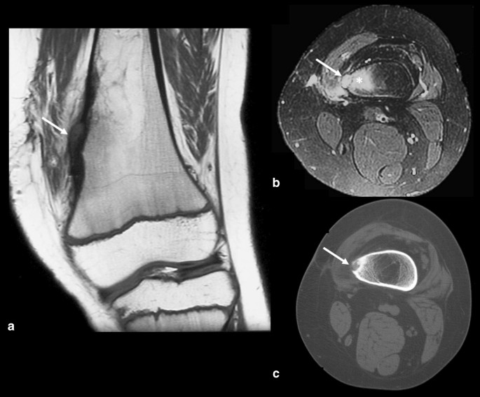
Radiologic Diagnosis of Osteoid Osteoma: From Simple to Challenging  Findings | RadioGraphics
Radiologic Diagnosis of Osteoid Osteoma: From Simple to Challenging Findings | RadioGraphics

Osteoid osteoma of the hip: imaging features | SpringerLink

Figure2.a: Magnetic resonance imaging (MRI) (T2WI) shows higher... |  Download Scientific Diagram
Figure2.a: Magnetic resonance imaging (MRI) (T2WI) shows higher... | Download Scientific Diagram

Clinical Vignette 8 | MIPHIDIC

Osteoid osteoma in an 18-year-old male patient presenting with pain in... |  Download Scientific Diagram
Osteoid osteoma in an 18-year-old male patient presenting with pain in... | Download Scientific Diagram

Osteoid Osteoma with a Multicentric Nidus: Interstitial Laser Ablation  under MRI Guidance
Osteoid Osteoma with a Multicentric Nidus: Interstitial Laser Ablation under MRI Guidance

Osteomyelitis (Suspected Acute)

Radiologic Diagnosis of Osteoid Osteoma: From Simple to Challenging  Findings | RadioGraphics
Radiologic Diagnosis of Osteoid Osteoma: From Simple to Challenging Findings | RadioGraphics

Postinterventional MRI findings following MRI-guided laser ablation of  osteoid osteoma - ScienceDirect
Postinterventional MRI findings following MRI-guided laser ablation of osteoid osteoma - ScienceDirect

Osteoid osteoma: the great mimicker | Insights into Imaging | Full Text

MR of osteoid osteoma. Sagittal T2-w fat saturated MRI image of a 1-cm... |  Download Scientific Diagram
MR of osteoid osteoma. Sagittal T2-w fat saturated MRI image of a 1-cm... | Download Scientific Diagram